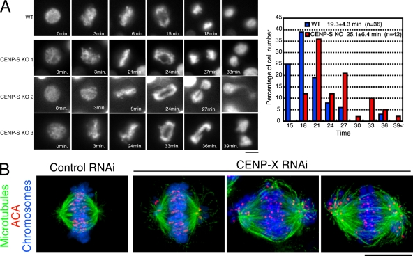
Figure 2.
Both CENP-S– and CENP-X–deficient DT40 cells are viable but show defects in mitotic progression. (A, left) Dynamics of chromosomes in wild-type (WT) or CENP-S–deficient cells visualized by time-lapse observation of living cells. Selected images of chromosomes from prophase to anaphase in these cells are shown. (right) Quantification of the time for progression from prophase to anaphase in wild-type and CENP-S–deficient cells as determined by time-lapse microscopy of living cells. (B) Chromosome morphology and α-tubulin staining (green) in human HeLa cells after siRNA-based knockdown for CENP-X. Human anticentromere antibodies (ACA) were used to detect the position of centromeres (red), and DNA (blue) was stained with Hoechst. Bars, 10 µm.

