
Figure 2. Progressive loss of proliferation markers in external granule layer.
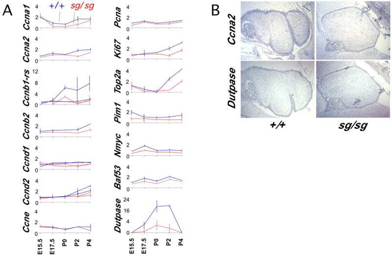
(A) Normalized expression data from Affymetrix Mu11K microarrays are plotted as line graphs. Staggerer samples are in red, littermate controls in blue. Average difference values for each probe set are normalized to make the average of all 24 hybridizations equal to 1. Horizontal lines indicate range of values among replicate samples. Multiple lines in a given plot represent overlay of normalized data from independent probe sets on the array. During this perinatal window, only the cells in the external granule layer should contribute in large number to pool of dividing cells in the cerebellum.
(B) Paired serial sections from control and staggerer specimens were mounted together on single slides and processed for in situ hybridization. In situ hybridization shows Ccna2 and dUTPase RNA expression is restricted to the EGL within the cerebellum at P2. Note the thinning of the staggerer EGL by this time.