Figure 2.
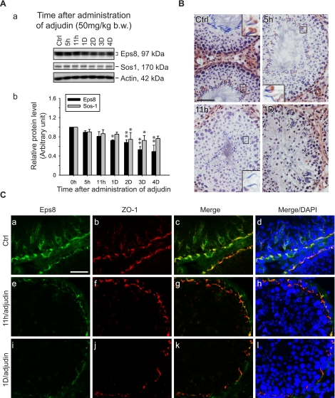
Changes in the steady-state level of Eps8 and its cellular localization in the seminiferous epithelium during adjudin-induced germ cell loss. Adult rats were treated with adjudin to induce germ cell loss resulting from anchoring junction restructuring in the seminiferous epithelium. A) a) Immunoblots of Eps8 and Sos1 in testis lysates following adjudin treatment, with actin as a loading control. b) Histogram summarizing results after normalizing each data point against actin. Protein levels at 0 h were arbitrarily set as 1. Bars are means ± sd; n = 3. *P < 0.05, **P < 0.01; 1-way ANOVA followed by Dunnett’s test. B) Immunohistochemistry of Eps8 using frozen testis sections after adjudin treatment. In control (Ctrl), Eps8 staining is clearly visible at the apical ES in most elongated spermatid heads in a stage VII tubule. The staining gradually weakens when elongated spermatids deplete from the epithelium at 5 and 11 h after adjudin treatment in non-stage VIII tubules. By 1 d, Eps8 is also diminished at the BTB site in stage V–VI tubules. Insets: magnified view of boxed area in same panel. C) Colocalization of Eps8 (green) and ZO-1 (red) in frozen testis sections after adjudin treatment. Eps8 at the BTB is much weakened at 11 h and 1 d after adjudin treatment, whereas ZO-1 remains relatively unchanged. Nuclei stained with DAPI (blue) illustrate germ cell loss from the epithelium. Scale bars = 80 μm (B); 40 μm (C).

