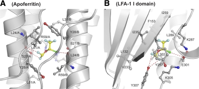
Figure 3.
Comparison of isoflurane-binding sites in apoferritin and I domain. Ribbon diagram showing isoflurane-binding sites in apoferritin (A) and LFA-1 I domain (B). Isoflurane-interacting residue side chains are shown as a ball-and-stick model with red oxygen and blue nitrogen atoms. L24/A and L24/B main chains are also shown as a ball-and-stick model (A). Isoflurane is shown as a ball-and-stick model with yellow carbon, red oxygen, green chloride, and sky blue fluorine atoms. Polar interactions of isoflurane with proteins are represented by red dashed lines.

