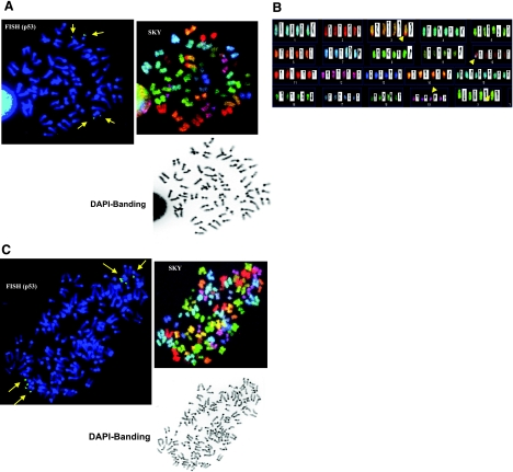
Figure 1.
Stable Aurora-B induces PCS in NmuMG cells, in both 2N and 4N cells. A) Mitosis carrying 2N range of chromosomes (42, XX with trisomies of no. 15 and 19), which show the phenomenon of total PCS, characterized by the separated and splayed chromatids and a discernable centromere involving all chromosomes (also known as C-anaphase cells). This is demonstrated by FISH, using BAC RP23-51O13 for p53 status (top left), SKY (top middle), and DAPI banding (top right). Because of the mitotic event leading to PCS, each chromatid of no. 11 chromosome is recognized to carry an individual FISH signal of p53. B) As clearly shown by SKY (bottom panel), the total number of chromatids is 84. C) Mitosis carrying 4N range of chromosomes (84, XXXX), as demonstrated by FISH using BAC RP23-51O13 for p53 status (top left), also showed PCS in all chromosomes, SKY (upper right) and DAPI banding (bottom left). Total number of p53 signals (i.e., 8) is demonstrated to coincide with the total number of chromatids of no. 11 chromosome, which clearly show total PCS events. Long arrows indicate p53 gene location. Chromosomal rearrangements were also noted in the SKY analysis, as indicated by short arrows in A, bottom panel.

