Figure 3.
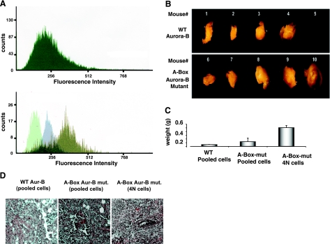
In vivo tumorigenicity of NmuMG cells stably expressing wild-type Aurora-B or Aurora-B mutant. A) Ploidy analysis of NmuMG cells, A-box-Aurora-B mutant stable transfectant before (top) and after sorting (bottom) by FACS. In the latter case, an equal number of the sorted cells was subjected to FACS analysis. Histograms are representative of 3 independent experiments. Histograms of sorted cells with 2N, 4N, and 8N DNA content are superimposed. Overlapping regions (dark green) represent cells with ploidy content other than that indicated. B) Nude mice (nu/nu) were subcutaneously injected into mammary fat pad with pooled cells overexpressing wild-type Aurora-B (top panel) or stable A-Box-Aurora-B mutant (bottom panel) or sorted tetraploid cells sorted as in panel A (tumors not depicted) at a concentration of 2 × 10⁶ cells/mouse (∼27 g total weight). Tumors were surgically removed after 8–10 wk postinjection and visualized using a Zeiss Stemi SV6 dissecting microscope, as described in Materials and Methods. Mouse 5 did not develop a tumor. C) Average tumor weights (g). Error bars = sd. Values of P < 0.05 indicate a statistically significant difference between groups using 1-way ANOVA test. D) Tumor histology shows random and blinded H&E-stained paraffin-embedded tissue sections that were sent to pathology for independent interpretation. Tumors from 4N cells show grade 3 adenocarcinoma, poorly differentiated neoplastic epithelial cells with significant nuclear atypical and high mitotic activities. There are irregular borders and evidence of tumor invading the adjacent layers.

