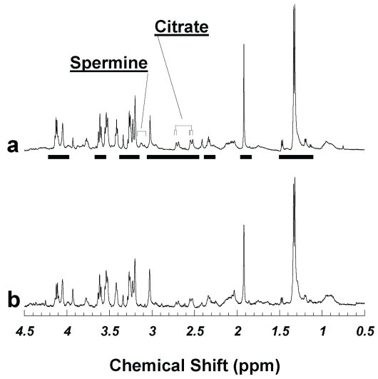
Figure 1.
Visually undifferentiated human prostate tissue HRMAS proton spectra from two cuts of the same surgical specimen of a cancerous prostate measured (a) in 2005 after being stored at −80 °C for 32 months, and (b) in 2002 when the sample was thawed after being frozen overnight. Quantitative pathology detected no histopathologically identifiable cancerous glands in either sample; other than stromal cells, the majority of prostate pathology in both samples was histopathologically benign epithelia, which comprised 46.1 and 33.8%, for (a) and (b), respectively. Figure 1 (b) was adopted from Figure 1 (b) of Ref. (Wu et al. 2003). Metabolite intensities analyzed in the current study are labeled with horizontal bars under spectrum 1a.

