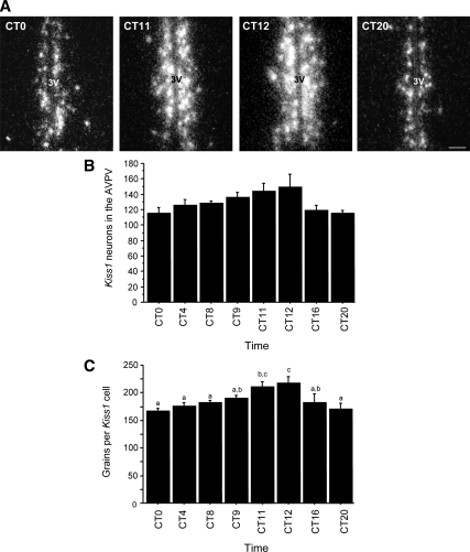
Figure 3.
A, Representative dark-field photomicrographs showing Kiss1 mRNA-expressing cells (as reflected by the presence of white clusters of silver grains) in the AVPV of OVX, E2-treated female mice that were housed in constant conditions and killed at different times throughout the circadian day. 3V, Third ventricle. B, Mean (±sem) number of Kiss1-expressing cells in the AVPV across the circadian day displayed a trend (P < 0.09) for more Kiss1 neurons during the late subjective afternoon/early evening. C, The amount of Kiss1 mRNA per cell, as indicated by the number of silver grains per cell, was significantly different across circadian time points, with highest values at CT 11 and CT 12 (P < 0.01); values with different letters differ significantly from each other. n = 4–6 animals per group.

