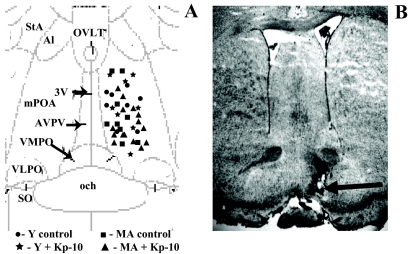
Figure 1.
Illustration of microdialysis probe placements in the medial preoptic area. A, The diagram corresponds to a coronal section at approximately 0.0 mm relative to Bregma (plate 33) in the atlas of Paxinos and Watson (81). 3V, Third ventricle; och, optic chiasm; VMPO, ventromedial preoptic nucleus; VLPO, ventrolateral preoptic nucleus; SO, supraoptic nucleus; Al, alar nucleus; StA, strial part preoptic nucleus; MA, middle-aged rats; Y, young rats; OVLT, organum vasculosum laminae terminalis. B, Photomicrograph of thionin-stained coronal section showing a representative probe placement between plates 32 and 33 by Paxinos and Watson (81). Magnification, ×40, shows the approximate location of a microdialysis probe. The arrow indicates the site of probe tip.

