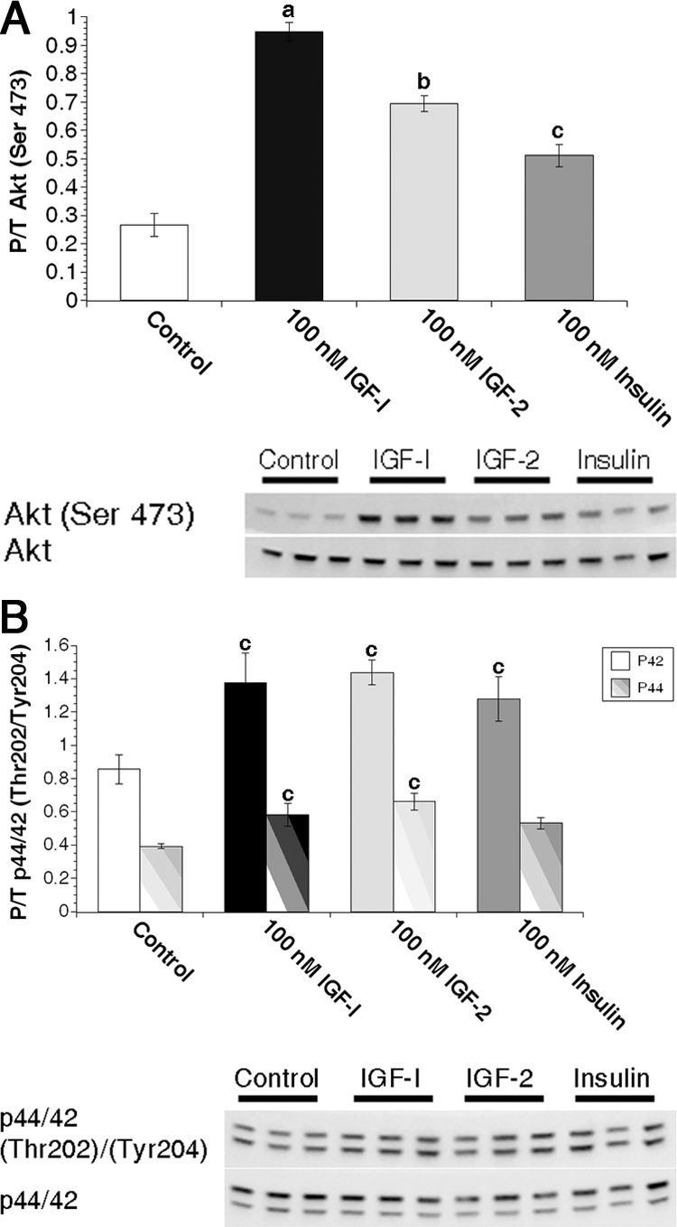
Figure 2.

IGF ligands stimulate Akt phosphorylation more efficiently than insulin in primary MECs. Freshly isolated primary MECs from virgin animals were treated with 0 nm (control) or 100 nm IGF-I, IGF-II, or insulin for 15 min immediately followed by protein isolation. A, Graphs depict densitometric analysis of Akt Western immunoblot images. Bars indicate mean ± sem of phosphorylated Akt (Ser473) adjusted to total Akt expression. B, Graphs depict densitometric analysis of p44/p42 Western immunoblot images. Bars indicate mean ± sem of phosphorylated p42 (Tyr204, solid bars) or p44 (Thr202, striped bars) adjusted to total protein. a, P ≤ 0.001 vs. control, IGF-II, and insulin; b, P < 0.01 vs. control and insulin; c, P ≤ 0.04 vs. control. n = 3 samples per treatment.
