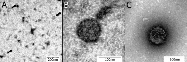
Figure 4.
Transmission electron microscopy images. A) Brain homogenate from an ABV4(+) PDD(+) African grey parrot (the inoculum used in this study). Three spherical virus-like particles approximately 60 nm in diameter are shown [arrows] (negative staining with uranyl acetate). B) A virus-like particle from the same specimen in "A" shown at greater magnification. This particle is 98 nm in diameter (negative staining with uranyl acetate). C) A virus-like particle from the brain of cockatiel 1. This particle is 99 nm in diameter and is showing bold projections on its circumference (negative staining with uranyl acetate).

