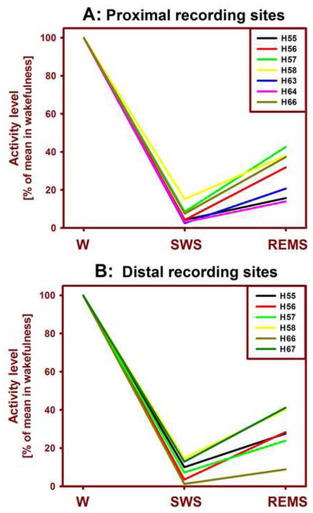
Figure 3.
Mean levels of lingual EMG across the three behavioral states in individual animals. The lingual EMG was lowest during SWS and then increased during REMS for both proximal (A) and distal (B) recording sites. Activity levels in SWS and REMS were normalized by their average levels during wakefulness (W). The levels of lingual EMG during SWS and REMS did not differ between the proximal and distal recording sites. Numbers preceded by “H” denote subjects (cf. Fig. 2).

