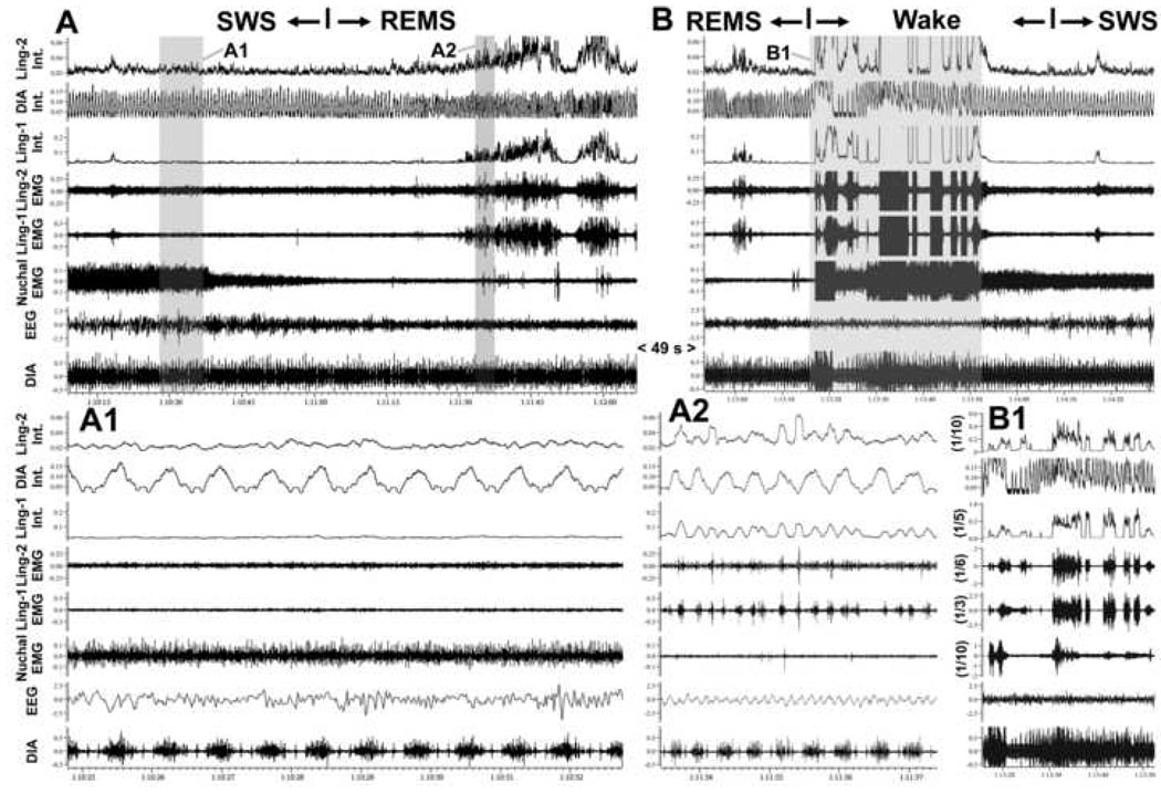
Figure 5.
Absence of respiratory modulation of lingual EMG during state transitions. A and B show two successive segments of EMG records from two intermediate sites in the tongue (Ling-1 and Ling-2; sites labeled in Fig. 2 as H65) and the diaphragm (DIA), as well as their integrated (Int.) versions, together with the cortical EEG and nuchal EMG taken from periods of state transitions. In the 113.5 s-long record shown in A, a transition from SWS to REMS was identified at the mark based on the atonia of nuchal EMG and appearance of theta rhythm in the EEG. In the 95.5 s-long record shown in B, awakening from REMS occurred and then the animal entered SWS. During the 49 s period separating A and B, REMS continued uninterrupted. The records in A1 (7.95 s) and A2 (3.95 s) show on an expanded time scale the shaded parts of the record in A. They correspond to the periods of SWS and REMS, respectively. A careful inspection of the raw and integrated diaphragmatic and lingual EMGs reveals no evidence of respiratory modulation of lingual EMG in A (Ling-1 is atonic and Ling-2 nearly atonic). In B, both lingual EMGs exhibit rhythmic, or semi-rhythmic bursts, but the phase and frequency of these rhythms is different from the respiratory rhythm in the diaphragm. B1 shows the 36.7 s-long, highlighted portion of the record in B using the same time scale as in B, but with the gains for lingual and nuchal EMGs reduced 3–10 times to show the full magnitude of activation present in these muscles around the time of awakening.

