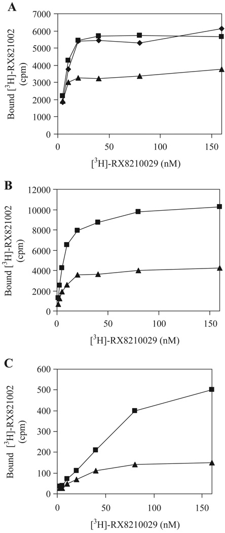
Fig. 4.
Effect of α2B-ARm on the transport of α2A-AR and α2C-AR. A. DDT-MF2 cells stably expressing α2A-AR were cultured on 100-mm dishes and transfected with 10 µg of pEGFP-N1 vector (squares), α2B-ARm-GFP (triangles) or AT1Rm-GFP (diamonds). B. HEK293T cells were co-transfected with 4 µg of α2A-AR in pcDNA3 and 6 µg of pEGFP-N1 vector (squares) or α2B-ARm-GFP (triangles). C. HEK293T cells were co-transfected with 4 µg of α2C-AR in pcDNA3 and 6 µg of pEGFP-N1 vector (squares) or α2B-ARm-GFP (triangles). Membrane preparation (25 µg) was incubated with increasing concentrations of [3H]-RX-821002 (1.25–160 nM) for 30 min. Specific binding was determined in duplicate and nonspecific binding determined in the presence of 10 µM rauwolscine as described under “Experimental Procedures”. The data shown are representative of three separate experiments.

