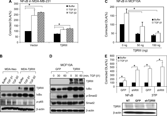
Fig. 2.
TβRIII negatively regulates NFκB signaling. (A and C) MCF10A and MDA-MB-231 cells were transfected with pNFκB-Luc, pRL-SV40 and TβRIII (300 ng per well in a 24-well plate for MDA-MB-231 cells, indicated amounts for MCF10A cells) using Fugene 6 and grown for 24 h. Cells were treated with buffer, TGF-β1 (100 pM) or TGF-β2 (200 pM) for 24 h and harvested for luciferase activity assay. Data are expressed as means ± SDs of at least three independent experiments. Statistical significance of differences was assessed using unpaired Student's t-tests (*P < 0.01). (B) MDA-MB-231-Neo and MDA-MB-231-TβRIII cells were treated with TGF-β1 or TGF-β2 for 1 h, harvested and subjected to western blotting. (D) Subconfluent MCF10A cells were infected with Adeno-TβRIII or GFP-adenovirus. After 2 days, cells were treated with TGF-β1 (200 pM) for the indicated times and harvested for western blotting with the indicated antibodies. The results shown are representative of at least three independent experiments. (E) MCF10A cells were infected with GFP or shRNA of TβRIII adenovirus and transfected with pNFκB and pRL-SV40 for reporter gene assay. After 1 day, cells were treated with TGF-β1 or TGF-β2 for 24 h and subjected to reporter gene assay and binding and cross-linking assay. Data are expressed as means ± SDs of at least three independent experiments. Statistical significance of differences was assessed using unpaired Student's t-tests (*P < 0.01).

