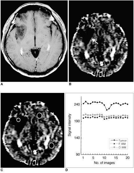
Fig. 3.
Case 19: Low-grade astrocytoma in a 41-year-old man.
A. Postcontrast T1-weighted image shows a non-enhancing low signal intensity tumor in the right basal ganglia.
B. Relative cerebral blood volume (rCBV) map shows low rCBV in the tumor (arrow).
C. rCBV map shows the placement of ROIs for measurement of rCBV in the tumor (small circle) and in contralateral frontal and occipital white matter (large circles).
D. Signal intensity-time curves measured at ROIs in C show less signal reduction in this tumor than in the high-grade gliomas seen in Figs. 1 and 2, suggesting that the vascularity of an astrocytoma is lower.

