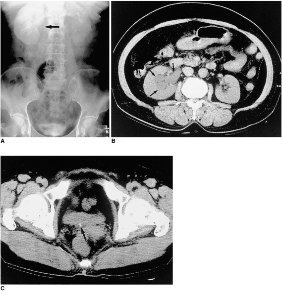
Fig. 3.
A 57-year-old woman with acute right flank pain.
A: EU; B, C: Transaxial UCT scans through the kidneys and bladder. A dense delayed nephrogram is seen in A, but the entire EU study revealed no stone shadow. On UCT a tiny stone at the right ureterovesical junction (arrow in C) is seen along with dilatation of the renal pelvocalyceal system (arrows in B).

