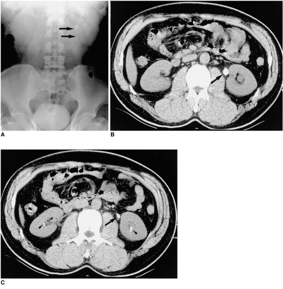
Fig. 4.
A 45-year-old male patient with acute flank pain.
A: EU; B, C: UCT. Two separate stones (arrows) are demonstrated in the left proximal ureter in A, but on UCT (B, C) they are seen as one elongated stone. UCT (C), however, demonstrates small renal stones (arrowheads), not seen in A, at the lower pole of the left kidney.

