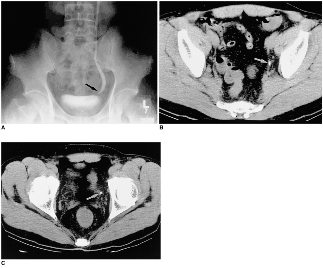
Fig. 5.
A 53-year-old man with acute flank pain.
A: EU; B, C: Two consecutive CT scans of the pelvis. A single stone is seen in the left distal ureter in A; UCT, however, clearly demonstrates two separate distal ureter stones (white arrows) in B and C. The smaller stone in the more distal ureter, seen in C, is obscured in A.

