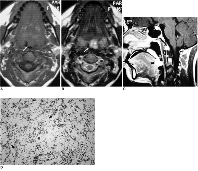
Fig. 2.
Schwannoma in a 16-year-old girl with swallowing difficulty.
A. T1-weighted axial image shows a well-defined, homogeneous mass (arrow), isointense to surrounding muscle, at the base of the tongue and encroaching on the airway.
B. T2-weighted axial image shows a mass with heterogeneous high signal intensity (arrow).
C. Enhanced T1-weighted sagittal image shows that the mass is located at the posterior one-third of the tongue, which corresponds to the region of the foramen cecum (black arrow). This mass significantly encroaches on the airway (white arrow).
D. Photomicrograph (original magnification ×40; H & E staining) shows spindle cells with some whirling and pallisading of their nuclei (arrows). The cells resemble Verocay bodies and enclose a space nearly devoid of nuclei (*). There is no evidence of malignancy.

