Fig. 3.
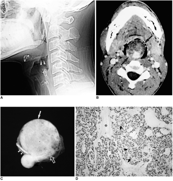
Myoepithelioma in a 35-year-old man with severe dyspnea and difficulty in swallowing.
A. Lateral view of plain cervical spine shows a lobulating, well-defined soft tissue density mass (arrow) at the base of the tongue, resting against the epiglottis (arrowheads) and encroaching on the airway.
B. Contrast-enhanced axial CT scan shows a well-defined mass with heterogeneous attenuation (arrows) at the base of the tongue. Since the anterior portion of this mass has low attenuation similar to that of subcutaneous fat, initial differential diagnosis included benign mature teratoma and benign minor salivary gland tumor such as pleomorphic adenoma.
C. Photograph of a cut section of gross specimen shows a well-demarcated yellowish mass (arrow) arising from the base of the tongue (open arrow). A lower small daughter nodule is covered by intact lingual mucosa. Note the intervening epiglottis (arrowheads) between the main mass and the nodule.
D. Photomicrograph (original magnification ×100; H & E staining) shows plasmacytoid-type myoepithelioma composed of small polygonal cells (arrows) with centrally located nuclei and abundant cytoplasm in a myxoid ground substance (*).

