Fig. 5.
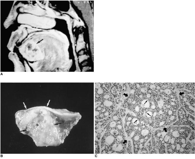
Adenoid cystic carcinoma in a 63-year-old woman with growing lingual mass.
A. Enhanced T1-weighted sagittal image shows a well demarcated, markedly enhancing mass in the submucosal area of the anterior tongue (arrow), with heterogeneous attenuation suggesting necrosis (*).
B. Photograph of a cut section of gross specimen shows a well-demarcated yellowish mass (black arrows) with a central irregular necrotic area (*) in the anterior region of the tongue. Note the presence of intact overlying lingual mucosa (white arrows).
C. Photomicrograph (original magnification ×100; H & E staining) shows tumor cells composed of uniform, small, angulated cells (thin arrows). Sharply defined cylindrical cores of hyaline material (*) create a cribriform, pseudocystic appearance (thick arrows), characteristic of grade-1 adenoid cystic carcinoma. Due to perineural invasion, the patient underwent hemiglossectomy and adjuvant chemotheraphy.

