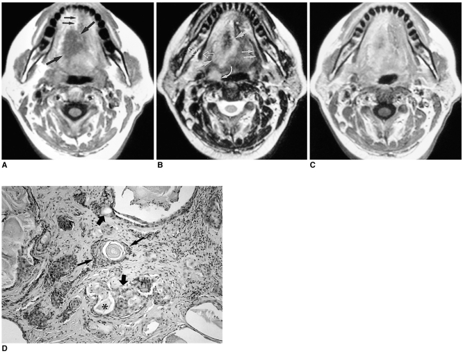
Fig. 6.
Mucoepidermoid carcinoma in a 48-year-old female with a painful tongue.
A. T1-weighted axial image shows a soft tissue mass with low signal intensity at the right lateral aspect of the tongue which extends across the midline (thick arrows). The lingual septum is seen only at the anterior half of the tongue (thin arrows). The right lateral margin of the mass is indistinct.
B. T2-weighted axial image shows a mass with increased signal intensity (thick arrows). Anterolateral extension of this mass is more easily recognized than on the T1-weighted image. The right lateral tissue plane and hyoglossus muscle are obliterated, though on the left side are visible (thin arrows). Note the prominent right palatine tonsil (curved arrow), suggesting tonsilar metastasis, and the intact right mylohyoid muscle (open arrows).
C. Enhanced T1-weighted axial image demonstrates heterogeneous, moderate enhancement, making differentiation of the mass from squamous cell carcinoma impossible.
D. Photomicrograph (original magnification ×100; H & E staining) demonstrates a mixture of the glandular component lined by well-differentiated cuboidal to columnar epithelium (thick arrows) containing abundant mucous material (*) and solid nests composed of squamous cells (thin arrows).

