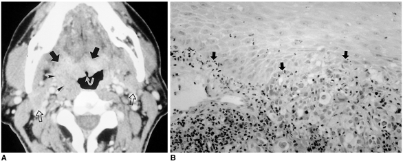
Fig. 9.
Metastasis from carcinoma of the bronchus in a 48-year-old man.
A. Contrast-enhanced axial CT scan shows an ill-defined, well-enhancing mass at the base of the tongue and right vallecula, with exophytic growth to the oropharyngeal cavity (arrows). The superficial portion of the mass is ulcerated (curved arrow). The mass extends laterally, causing prominence of the right palatine tonsil (arrowheads). Multiple bilaterally enlarged lymph nodes can also be seen (open arrows).
B. Photomicrograph (original magnification ×200; H & E staining) demonstrates poorly differentiated adenocarcinoma (arrows) metastasized from bronchogenic carcinoma. Palliative radiotherapy was performed.

