Abstract
We present the characteristic plain radiographic and intravenous urographic (IVU) findings of calculus formed over a hair. A 66-year-old man who had been quadriplegic for 40 years because of vertebral injury was admitted for further evaluation of frequent urinary tract infection. Plain radiography showed a linear, serpiginous calcification in the lower abdomen, and IVU revealed a round filling defect with linear radiopacity in the bladder, suggesting calculus. The gross appearance of the stone after extraction demonstrated that calcification had formed over a hair.
Keywords: Urolithiasis, Bladder stone, Hair, Radiography
Bladder calculi occur often in patients with urinary stasis and infection, and a foreign body in the bladder may serve as a nidus for stone formation. Reports have shown that a multitude of foreign bodies are capable of acting as a center for bladder calculi, but a hair, introduced inadvertently into the bladder during catheterization, has rarely been reported as the cause of stone formation.
We present the characteristic plain radiographic and intravenous urographic (IVU) findings of calculus over a hair introduced during catheterization.
CASE REPORT
A 66-year-old man who had been quadriplegic for 40 years because of cervical vertebral injury during the Korean War was admitted for further evaluation of frequent urinary tract infection. Urinalysis revealed numerous bacteria and white blood cells.
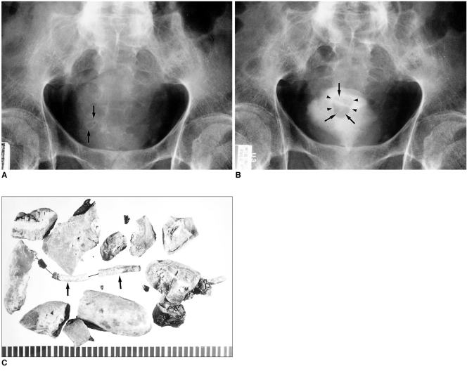
Plain radiography showed a linear, serpiginous calcification in the lower abdomen (Fig. 1A), and IVU demonstrated a round filling defect with linear radiopacity in the bladder, suggesting calculus (Fig. 1B).
Fig. 1.
66-year-old man with bladder calculus formed over a hair nidus.
A. Plain radiograph shows serpiginous radiopacity in the lower abdomen (arrows).
B. Intravenous urograph demonstrates round filling defect with linear radiopacity (arrows) in the bladder (arrowheads).
C. Gross appearance of the calculus after extraction shows that serpentine calcification had formed over a hair (arrows).
Ultrasonic lithotripsy was performed, and using forceps, several stone fragments were removed. The results of analysis showed that they contained a mixture of calcium oxalate, magnesium, ammonium and carbonate. The gross appearance of the stone after extraction demonstrated that calcification had formed over a hair (Fig. 1C).
DISCUSSION
For a variety of reasons, including immobilization hypercalciuria, urinary stasis, infection, and the introduction of a Foley catheter, patients with spinal cord injury are predisposed to calculi (1).
Calculus formation over a hair introduced into the bladder is a preventable complication (2). Being a foreign material, hair in this location is an ideal site for crystalline precipitation, which may then be perpetuated by the reaction of urothelium with the extraneous material (3).
The absence of normal micturition in spinal cord injury patients exacerbates the problem because it prevents spontaneous passage of small stone fragments. Some therefore undergo intermittent urethral catherization, and in some care centers it is routine practice to recommend periodic shaving of pubic hair in such patients (4).
In 1983, Amendola et al. reported that during a 12 - year period, plain radiographs of three of eight patients with bladder calculi formed over a hair nidus showed characteristic serpiginous, linear calculi (1). To the best of our knowledge, however, the IVU findings in patients with bladder calculi over a hair nidus have not been previously reported.
In our case, IVU revealed serpiginous radiopacity within the filling defect. Compared with the pathologic specimen, the serpiginous dense radiopacity seen at plain radiography and IVU revealed a calculus formed over a hair.
When plain radiographs or IVU in patients with a neurogenic bladder show serpiginous radiopacity, the presence of bladder calculi formed over hair introduced into the bladder during catheterization may be suspected. An understanding of this etiology may help prevent bladder calculus.
References
- 1.Amendola MA, Sonda LP, Diokno AC, Vidyasagar M. Bladder calculi complicating intermittent clean catheterization. AJR. 1983;141:751–753. doi: 10.2214/ajr.141.4.751. [DOI] [PubMed] [Google Scholar]
- 2.Solomon MH, Koff SA, Diokno AC. Bladder calculi complicating intermittent catheterization. J Urol. 1980;124:140–141. doi: 10.1016/s0022-5347(17)55333-1. [DOI] [PubMed] [Google Scholar]
- 3.Derry F, Nuseibeh I. Vesical calculi formed over a hair nidus. Br J Urol. 1997;80(6):965. doi: 10.1046/j.1464-410x.1997.00340.x. [DOI] [PubMed] [Google Scholar]
- 4.Vaidyanathan S, Bingley J, Soni BM, Krishnan KR. Hair as the nidus of a bladder stone in a traumatic paraplegic patient. Spinal Cord. 1997;35(8):558. doi: 10.1038/sj.sc.3100426. [DOI] [PubMed] [Google Scholar]


