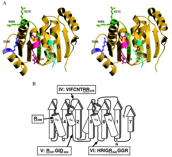
Figure 1.
Structure of the carboxyl-terminal domain of eIF4A. (A) Stereoview, ribbon drawing of the structure. Conserved motifs are colored as follows: motif IV, VIFCNTRR, residues 263–270, green; “conserved R” motif, residue Arg-298, purple; motif V, RGID, residues 321–324, magenta; motif VI, HRIGRGGR, residues 345–352, cyan. The strands of the β-sheet are labeled sequentially. This and subsequent ribbon drawings were prepared with molscript (34) and rendered with raster3d (35). (B) Topology diagram of the structure. β-Strands are shown as arrows; α-helices, as cylinders. β-Strands and α-helices are labeled sequentially as 1–7 and α1– α6, respectively. Sequences of the conserved motifs are shown in boxes; residues whose side chains are illustrated in A and subsequent figures are underlined.

