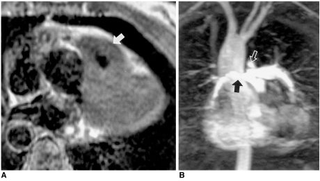
Fig. 5.
Tetralogy of Fallot in a 37-year-old female. MRI rather than catheter angiography was performed prior to surgery.
A. Axial spin-echo MR image shows severe infundibular stenosis (arrow).
B. Three-dimensional contrast-enhanced MR angiography shows small right pulmonary artery (solid arrow) and a ductus diverticulum (open arrow). Note, too, the right-sided aortic arch with mirror image branching.

