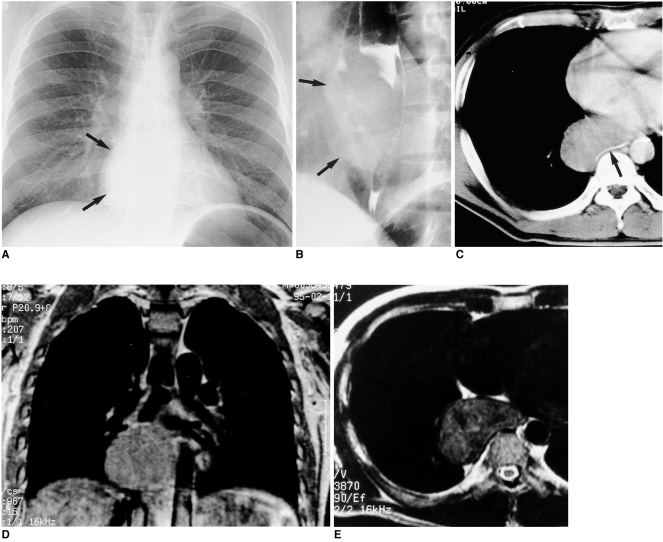
Fig. 1.
Esophageal leiomyoma in a 35-year-old man who had suffered substernal pain for two months.
A. Chest radiography reveals a right retrocardiac soft-tissue mass (arrows), with obliteration of the azygoesophageal recess interface.
B. Esophagography indicates the presence of a large, smoothly elevated filling defect in the distal esophagus. Also note the extraluminal component of the mass (arrows).
C. Enhanced CT scan (10-mm collimation) obtained at the ventricular level shows a homogeneous, iso-attenuated, pear-shaped mass, 75×45 mm in diameter, in the right azygoesophageal recess. Note the presence of gastrograffin-filled esophageal lumen (arrow).
D. Enhanced coronal T1-weighted MR image reveals a slightly hyperintense lesion in the azygoesophageal recess area.
E. T2-weighted MR image obtained at a level similar to that in C reveals a lesion slightly more hyperintense than chest wall muscle.

