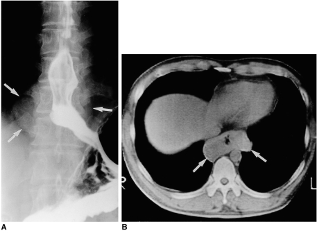
Fig. 3.
Esophageal leiomyoma in a 37-year-old man with mild dysphagia.
A. Esophagography shows a soft tissue mass (arrows) surrounding the opacified esophageal lumen. Note that there is mild passage disturbance and mucosal rigidity in the distal esophagus.
B. Unenhanced (10-mm collimation) CT scan obtained at another hospital at the level of the liver dome reveals a soft tissue mass measuring 80×40 mm (arrows) surrounding the gas-filled esophageal lumen.

