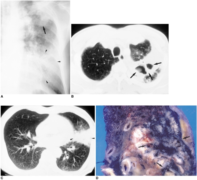
Fig. 4.
60-year-old male who presented with hoarseness.
A. Initial chest radiograph shows consolidation (arrow) in the left upper lung zone and ill-defined ground-glass opacity (arrowheads) in the left lower lung zone. Because acid-fast bacilli were present in sputum, the patient underwent anti-tuberculous chemotherapy.
B, C. CT scans obtained two months after A, due to persistent symptoms, show cavitary lesions (arrows) in the apicoposterior segment and segmental consolidation (arrowheads) in the lingular division of the left upper lobe.
D. Bronchoscopy demonstrated adenocarcinoma in the lingular division. In the pathologic specimen, a pinkish tumor, which proved to be tuberculous granuloma, engulfed the pigmented area (arrows).

