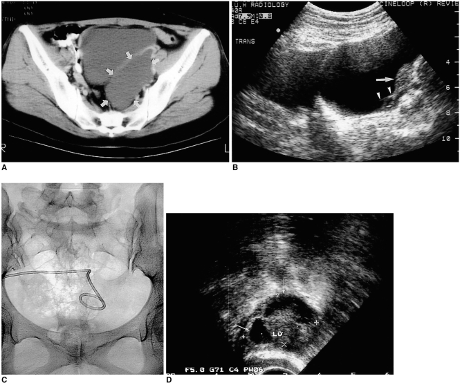
Fig. 1.
A 37-year-old woman who underwent total abdominal hysterectomy for uterine leiomyoma ten years earlier presented with lower abdominal pain (Patient 1).
A. Enhanced CT shows an elongated cystic mass with no solid component on the left side of the pelvis (arrows).
B. Transabdominal ultrasonogram of the pelvis in the transverse plane indicates a persistent cystic mass with internal septation (arrow-heads) after simple aspiration. The left ovary may be observed (arrow).
C. An 8.5-Fr pigtail catheter was introduced into the lesion transabdominally, and povidone-iodine was used for sclerotherapy.
D. Transvaginal ultrasonogram obtained 54 months after the procedure shows scanty fluid (arrows) around the left ovary (LO).

