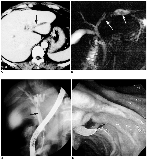
Fig. 1.
Biliary ascariasis in a 44-year-old woman.
A. Upper abdominal CT scan shows dilation of the intrahepatic bile ducts in the left lobe and a slightly hyperattenuating lesion (arrow) within the dilated bile duct.
B. MR cholangiogram shows a linear, tubular hypointense filling defect (white arrows) in the dilated bile duct.
C. Endoscopic retrograde cholangiogram shows a linear filling defect (arrow) in the left intrahepatic and common bile duct.
D. Photograph obtained during endoscopic extraction of the worm (arrow) shows that this is whitish and extends through the papilla of Vater.

