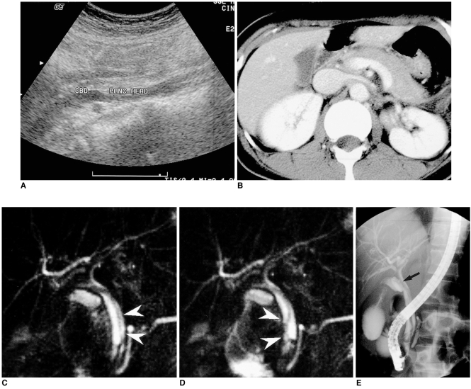
Fig. 2.
Biliary ascariasis in a 41-year-old woman.
A. Upper abdominal ultrasonogram shows mild dilatation of the common bile duct.
B. Contrast-enhanced upper abdominal CT scan shows mild diffuse enlargement of the pancreas.
C. MR cholangiogram shows a hypointense tubular filling defect (arrowheads) in the common bile duct.
D. Subsequent MR cholangiogram obtained 5 seconds after C demonstrates movement of the worm in the dilated bile duct.
E. Endoscopic retrograde cholangiogram shows a linear filling defect (arrow) in the contrast-filled common bile duct.

