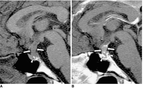
Fig. 11.
Pituitary adenoma in a 42-year-old man.
A. Sagittal T1-weighted MR image (600/15) shows marked thickening of the pituitary stalk (arrows). The posterior lobe, displaced by the lesion, is seen as several foci of high signal intensity (arrowheads).
B. Contrast-enhanced sagittal T1-weighted MR image (600/15) demonstrates homogeneous mild enhancement of the thickened pituitary stalk, which extends into the pituitary fossa and displaces the normal pituitary gland to the left side. Due to strong enhancement, the displaced and compressed pituitary gland is seen as multiple foci of high signal intensity (arrows). Although the mass was found to completely encircle the pituitary stalk, at surgery the two were separated easily. Pituitary adenoma was pathologically proven.

