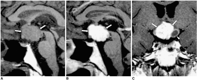
Fig. 12.
Craniopharyngioma in a 34-year-old man.
A. Sagittal T1-weighted MR image (600/15) shows a large lobulated mass (arrows) involving the suprasellar area and hypothalamus. The posterior lobe no longer shows high signal intensity.
B, C. Contrast-enhanced sagittal (B) and coronal (C) T1-weighted MR images (600/15) show strong enhancement of the solid component of the tumor (arrows), which also has a smaller cystic component (arrowhead in C). Craniopharyngioma was pathologically proven.

