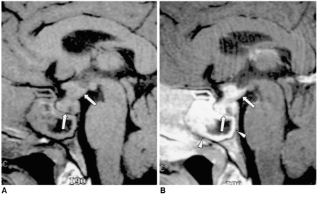
Fig. 6.
Churg-Strauss syndrome in a 38-year-old man.
A. Sagittal T1-weighted MR image (600/15) reveals an isointense mass involving the hypothalamus, pituitary stalk and pituitary gland (arrows). The posterior lobe no longer shows high signal intensity. Prominent mucosal thickening of the sphenoid sinus is apparent.
B. Contrast-enhanced sagittal T1-weighted MR image (600/15) demonstrates strong enhancement of the mass (arrows), and there is a non-enhancing portion. The thickened mucosa of the sphenoid sinus is intensely enhanced, representing sphenoid sinusitis (arrowheads). Churg-Strauss syndrome was pathologically proven.

