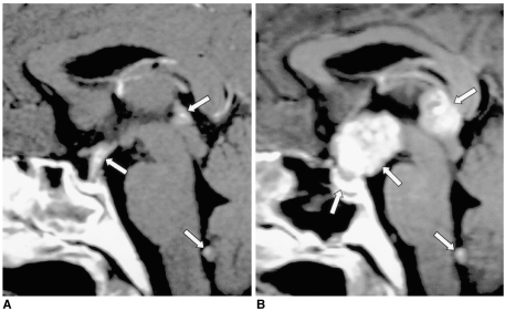
Fig. 7.
Germinoma in a 29-year-old man.
A. Initial contrast-enhanced sagittal T1-weighted MR image (600/15) depicts mild thickening and homogeneous enhancement of the pituitary stalk. In addition, small nodular enhancing lesions are seen in the pineal gland outlet of the fourth ventricle (arrows).
B. Contrast-enhanced sagittal T1-weighted MR image (480/14) obtained one year later, without treatment, shows a marked increase in the size and inhomogeneous enhancement of those lesions, and also of the pituitary gland (arrows). The focal enhancing lesion seen within the fourth ventricle appears to be cerebrospinal seeding. Germinoma was pathologically proven.

