Fig. 3.
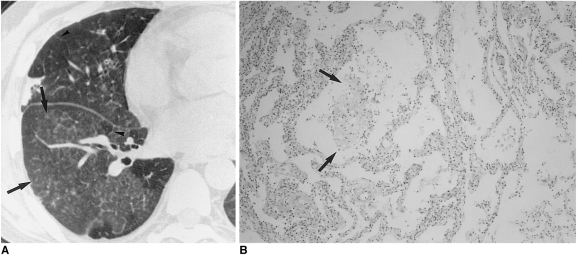
Cytomegalovirus pneumonia in a 30-year-old woman who presented with fever two months after kidney transplantation.
A. High-resolution (1.0-mm collimation) CT scan obtained at level of right inferior pulmonary vein shows mixed areas of ground-glass opacity and small poorly-defined centrilobular nodules (arrows). There is associated interlobular septal thickening (arrowheads).
B. Photomicrograph of transbronchial lung biopsy specimen obtained from superior segment of right lower lobe shows intra-alveolar collection of macrophages, fibrin and red blood cells (arrows) that corresponds to the centrilobular nodules seen on CT scans. There is associated alveolar wall thickening, with interstitial inflammatory cell infiltration (H & E, ×200).

