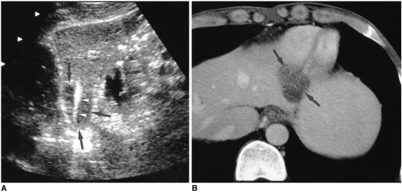
Fig. 3.
HCC in a 45-year-old man.
A. Longitudinal US scan of left hepatic lobe shows a 2.8-cm low echogenic HCC (arrows) which is fusiform in shape. The mass abuts on both liver capsules. Note the 3-cm active tip (arrowheads) of the straight cooled-tip electrode, which conforms to the shape of the mass. A single ablation lasted for 12 minutes.
B. Portal-phase CT scan obtained 30 minutes after RF ablation reveals the presence of an oval low-attenuating lesion (arrows), with absence of contrast enhancement within the ablated area. This indicates complete necrosis.

