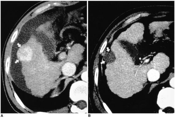
Fig. 4.
HCC in a 57-year-old man.
A. Hepatic arterial-phase CT scan depicts a 4-cm HCC (arrows) in the subcapsular area of the right hepatic lobe, which shows an outward bulge. Using a 3-cm expandable needle electrode, four ablations were performed. The deep portion of the mass was treated first, and then the exophytic portion.
B. Portal-phase CT scan obtained 13 months after RF ablation shows an ablated lesion (arrows) with no contrast enhancement, indicating complete necrosis.

