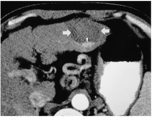
Fig. 7.
Hepatic arterial-phase CT scan obtained one month after RF ablation of a 4-cm HCC in the left hepatic lobe reveals the presence of a round low-attenuating lesion (arrows). Note the focal contrast enhancement (arrowhead) in the posterior aspect of the ablated lesion, indicative of viable untreated tumor. Additional RF ablation was subsequently performed.

