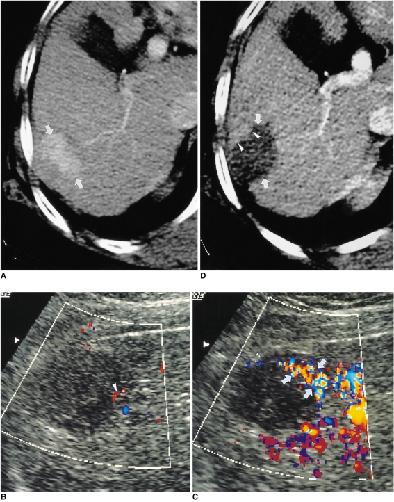
Fig. 9.
HCC in a 67-year-old woman.
A. Hepatic arterial-phase CT scan shows an oval enhancing HCC (arrows), 4 cm in its greatest diameter, at the periphery of the right hepatic lobe.
B. Unenhanced oblique power Doppler US scan obtained 18 hours after RF ablation shows a subtle flow signal (arrowhead) in the antero-superior aspect of the ablated lesion.
C. Contrast-enhanced oblique power Doppler US scan obtained immediately after B, above, shows peripheral flow signals (arrows) within the anterior aspect of the RF-ablated lesion, representing residual tumor. The residual flow signals seen on this contrast-enhanced scan are better appreciated than on nonenhanced power Doppler US scan.
D. Hepatic arterial-phase CT scan obtained 2 hours after RF ablation shows focal nodular contrast enhancement (arrowheads), indicative of residual tumor, in the anterior aspect of the ablated lesion (arrows). The enhanced area corresponds to that in which residual flow signals are present in the image described in C, above.

