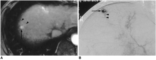
Fig. 2.
A 45-year-old man with small rapidly enhancing hemangioma in a cirrhotic liver resulting from chronic B-viral hepatitis.
A. Arterial dominant phase contrast-enhanced spoiled gradient-echo MR image (140/2.7) shows strong enhancement of a 0.9-cm tumor (arrow) with wedge-shaped temporal peritumoral enhancement (arrowheads). On unenhanced T1-and T2-weighted, and delayed contrast-enhanced MR images (not shown), the area of wedge-shaped enhancement could not be distinguished from surrounding hepatic parenchyma.
B. Capillary phase of hepatic arteriography shows near complete filling-in of the tumor by contrast agent (arrow), with opacification of a small, proximal portal vein branch (arrowheads), suggesting a transtumoral shunt or drainage of the hyperdynamic tumor by this branch.

