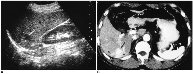
Fig. 10.
A 55-year-old woman with a hepatic hemangioma in the right lobe.
A. Sagittal US shows a well-defined hypoechoic mass (arrow).
B. Enhanced CT scan of the liver obtained during the hepatic arterial phase shows diffuse rapid enhancement of the tumor (arrow) with peritumoral wedge-shaped parenchymal enhancement (arrowheads), suggesting associated arterioportal shunt.

