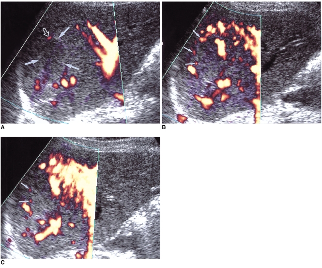
Fig. 11.
A 54-year-old woman with a hepatic hemangioma in the right lobe.
A. Unenhanced power Doppler US shows a hypoechoic mass with an echogenic border (arrows). The lesion shows minimal power Doppler signal in its periphery (open arrow) and optimized parameters (a pulse repetition frequency of 1,000 Hz and a medium wall filter).
B, C. Dynamic contrast-enhanced power Doppler US scans obtained 30 seconds (B) and 90 seconds (C) after the initiation of contrast injection show dot-like enhancement (small arrows) at the periphery of the mass. However, unlike centripetal fill-in enhancement, characteristic of hemangioma, the enhanced area revealed by power Doppler US is smaller 90 seconds after enhancement than at 30. Even with the use of microbubble agents, power Doppler US is, therefore, due to its insensitivity to slow flow, able to characterize hepatic hemangiomas to only a limited extent.

