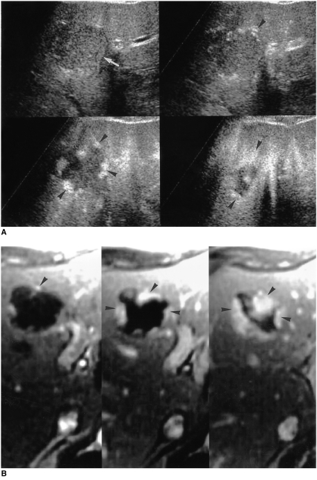
Fig. 12.
A 52-year-old man with two hepatic hemangiomas in the left lobe.
A. Pulse-inversion harmonic US scans obtained prior to contrast injection show a hypoechogenic hepatic hemangioma (arrow) in the medial segment of the left lobe. Serial contrast-enhanced US scans obtained 14, 62, and 139 seconds after injection show peripheral globular enhancement with progressive centripetal fill-in (arrowheads).
B. Serial dynamic contrast-enhanced T1-weighted MR images obtained immediately, 60, and 180 seconds after the administration of gadolinium-DTPA depict early peripheral nodular and globular enhancement with progressive centripetal fill-in (arrowheads), characteristic of hepatic hemangiomas. The enhanced areas seen on MR images are nearly identical to those seen on serial contrast-enhanced pulse-inversion harmonic US scans.

