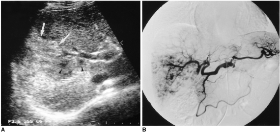
Fig. 5.
A 66-year-old man with a diffuse hemangioma in the right lobe.
A. Transverse US shows a large heterogeneous mass (arrows). The major portion of the lesion shows hyperechogenicity, especially in the periphery, and within it scattered hypoechoic foci are noted (arrowheads). Although the lesion abuts the right portal vein, there is no evidence of invasion of this vessel.
B. Celiac angiogram shows diffuse enhancement with scattered foci of contrast material puddling.

