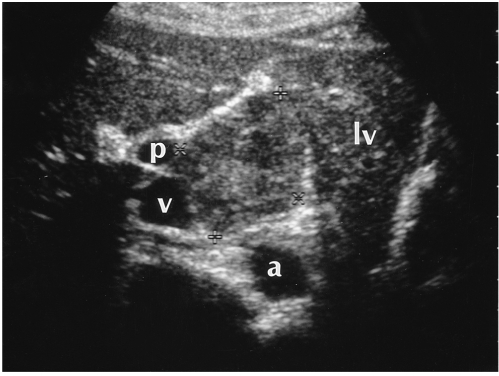
Fig. 7.
A 34-year-old woman with an exophytic hepatic hemangioma in the caudate lobe. Transverse US shows a large square-shaped mass surrounded by the left lobe of the liver (lv), aorta (a), inferior vena cava (v), and right portal vein (p). The lesion shows heterogeneous echogenicity and has a multiple internal hypoechoic portion.

