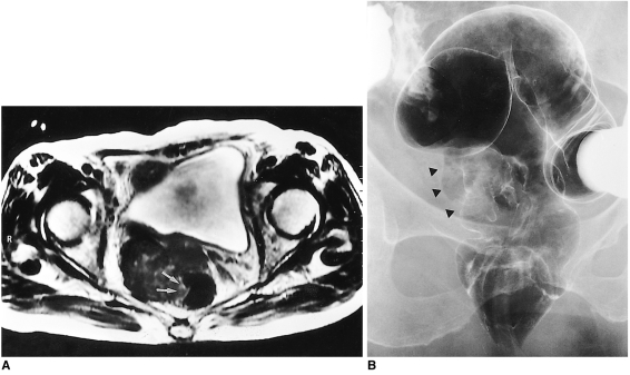
Fig. 3.
Recurrence at the primary site with posterior extension to the rectum in an 80-year-old woman who underwent radical hysterectomy.
A. T2-weighted axial MR image shows an irregular shaped, low signal mass between the bladder and rectum. The mass is continuous with the rectal wall (arrows), and protrudes into the rectal lumen.
B. Barium enema shows luminal irregularity and mucosal destruction of the lateral wall of the rectum, indicating the invasion of rectal mucosa (arrowheads).

