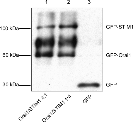
Figure 3. Western blotting of the expressed GFP-labelled Orai1 and STIM1.
Each lane was loaded with lysates (2 μg of protein) of HEK293 cells transfected with GFP-Orai1 and GFP-STIM1 containing plasmids at a molar ratio of 4 : 1 (lane 1); GFP-Orai1 and GFP-STIM1 containing plasmids at a molar ratio of 1 : 4 (lane 2); or GFP alone containing plasmid (lane 3). The blot was probed with anti-GFP antibody. The relative amounts of the expressed Orai1 and STIM1 proteins in the same transfection and between transfections were estimated by measuring optical densities of the corresponding bands (n= 3).

